Medical Thermal Imaging
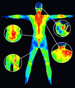
 Medical Thermal Imaging, also known as, Digital Infrared Thermal Imaging (D.I.T.T), or Thermography, is a non-invasive clinical imaging technique for detecting and monitoring a number of diseases and physical injuries by showing any thermal abnormalities present in the body. Thermography uses special infrared-sensitive cameras to digitally record images of the variations in surface temperature of the human body. The recorded images are calling thermograms. These thermograms are then analyzed by a medical doctor who will give diagnosis/prognosis based on the images.
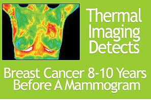
Breast Thermography is approved by the FDA as an alternative to a traditional mammogram, and can detect signs of cancer up to ten years earlier. As a safe, non-invasive screen technique for breast cancer, there is no touching or compression of the breast during the exam, which only takes minutes to complete.
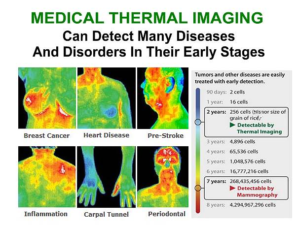
Thermography can be used as an aid in diagnosis and prognosis, as well as monitoring therapy progress for conditions and injuries which include: Artery Inflammation, Arthritis, Back Injuries, Breast Cancer, Breast Disease, Carpal Tunnel Syndrome, Cystic Formations, DCIS, Dental & TMJ, Digestive Disorders, Disc Disease, Diverticulitis, Fibromyalgia, Headache/Migraine Cause, Heart Disease, IBC, Inflammatory Pain, Nerve Damage, Periodontal Disease, Referred Pain Syndrome, RSD (CRPS), Scoliosis, Skin Cancer, Sprain/Strain, Stroke Screening, Thrombosis (Deep Vein), Unexplained Pain, Varicosities, Vascular Disease, Whiplash, and much more!
Benefits of Medical Thermal Imaging
(Thermal Imaging vs. Mammography)
- No intense pain, errors, or exposure to harmful radiation.
- Earlier detection: Generally, a tumor is first detected by a mammogram when it is about 2.5 cm, or the size of a dime, and at this stage it has been growing for at least 8 years. Thermography can detect cancer 8-10 years earlier than a traditional mammogram when it is in its earlier stages.
- Mammography can be a cause for multiple unnecessary biopsies. Studies have shown that biopsies for 70% to 80% of all positive mammograms do not show any presence of cancer. Breast Thermography has an average accuracy of 90%, and research shows that it can significantly improve long-term survival rates.
Most insurance companies cover 50% of a scan, but check with yours before booking and appointment.

Frequently Asked Questions
-
What is Thermography?Medical Thermal Imaging, also known as, Digital Infrared Thermal Imaging (D.I.T.T), or Thermography, is a non-invasive clinical imaging technique for detecting and monitoring a number of diseases and physical injuries by showing any thermal abnormalities present in the body. Thermography uses special infrared-sensitive cameras to digitally record images of the variations in surface temperature of the human body. The recorded images are calling thermograms. These thermograms are then analyzed by a medical doctor who will give diagnosis/prognosis based on the images.
-
Can thermal imaging, or Thermography, serve as an alternative to Mammography?
 Yes, the FDA has approved Thermography as a safe, non-invasive screening technique for breast cancer. There is no touching or compression of the breast during the exam, which only takes minutes to complete. It can also detect signs of cancer up to 10 years earlier without the exposure to X-Ray which is reported to actually increase the risk of cancer.
 -
Can I have a Thermal Breast Evaluation if I’ve had breast surgery (lumpectomy, mastectomy, breast implants/reduction)?Yes. Regardless of previous surgery to the breast tissue, your Thermal Imaging can be performed safely and accurately. In fact, mammography’s effectiveness is limited after such surgeries. There are guidelines as to when the Thermal Evaluation should be performed in relation to surgery.
-
What conditions can Thermography be used to diagnose?Thermography can be used for diagnosis and prognosis of many medical conditions:Altered Ambulatory Kinetics
Altered Biokinetics Brachial Plexus Injuries
Biomechanical Impropriety
Breast Disease
Bursitis
Inflammatory Disease
Int. Carotid Insufficiency
Infectious Disease
Ligament Tear
Lower Motor Neuron Disease
Lumbosacral Plexus Injury
Malingering
Median Nerve Neuropathy
Morton’s Neuroma
Muscle Tear
Musculoigamentous Spasm
Musculoigamentous Spasm
Myofascial Irritation
Nerve Entrapment
Nerve Impingement
Nerve Pressure
Tendonitis
TMJ DysfunctionCarpal Tunnel Syndrome
Compartment Syndrome
Cord Pain/Injury
Deep Vein Thrombosis
Disc Disease
Dystrophy
Facet Syndromes
Ext. Carotid Insufficiency
Nerve Root Irritation
Nerve Impingement
Nerve Stretch Injury
Neuropathy
Neurovascular Compression
Neuralgia
Neuritis
Neuropraxia
Neoplasia
Nutritional Disease
Periodontal Disease
Peripheral Axon Disease
Raynaud’s
Referred Pain Syndrome
Trigeminal Neuroalgia
Trigger PointsGrafts
Heart Disease
Hysteria
Headache Evaluation
Herniated Disc
Herniated Disc Pulposis
Hyperaesthesia
Hyperflexion Injury
Reflex Symp. Dystrophy
Ruptured Disc
Skin Cancer
Somatization Disorders
Soft Tissue Injury
Sprain/Strain
Stroke Screening
Synovitis
Sensory Loss
Sensory Nerve Abnormality
Skin Abnormalities
Somatic Abnormality
Superficial Vascular Disease
Temporal Arteritis
Ulnar Nerve Entrapment
Whiplash end edit
Contact Us
- 3551 Camino Mira Costa Suite CSan Clemente, CA 92672
- (949) 240-6713
- info@o2-wellness.com
- Get Directions
- Send Us A Message

